


Cloudy Eyes in Dogs: Why It Usually Isn't a Cataract, and How to Tell
Most cloudy-eyed dogs do not have cataracts at all. Here is how to tell a harmless aging change from a real cataract, and what each one actually calls for.

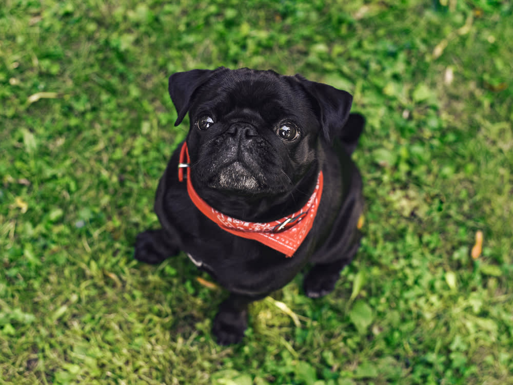
Here is the single most useful thing to know about a dog with a cloudy eye: most of the time, it is not a cataract at all. When owners come in worried that a cataract is forming, the far more common answer is a harmless aging change that needs nothing but a name. So before we get into cataracts, symptoms, and surgery, let’s start where a vet actually starts, with the question that decides everything: is this cloudiness the dangerous kind or the ordinary kind?
The look-alike that fools almost everyone
The condition most often mistaken for a cataract is nuclear sclerosis, and telling them apart is the whole game.
Nuclear sclerosis is a normal change in the lens that shows up in most dogs older than about six. Over the years the lens keeps making fibers but cannot shed the old ones, so the center grows denser and takes on a slight blue-gray haze. It usually appears in both eyes at once, does not meaningfully blur vision, and requires no treatment whatsoever. If your ten-year-old dog’s eyes have taken on a faint smoky tint but they still navigate the house and yard fine, sclerosis is the likely explanation.
A true cataract is different. It is an actual opacity in the lens, a cloudy film that physically blocks light from reaching the retina, and it can genuinely rob a dog of sight.
The tell is simple, and it is why this one belongs to your vet rather than a home guess.
The distinction sounds subtle, but it changes everything about what happens next. I shine a focused light into the eye. If I can see through the lens to the retina at the back, it is nuclear sclerosis and I reassure the owner. If the lens blocks my view of the retina, we are dealing with a real cataract and a real plan.
If it is sclerosis, you are done: no drops, no surgery, no worry. If it is a cataract, the rest of this guide is for you.
What a cataract actually is
The lens sits just behind the iris, the colored part of the eye, and its job is to focus incoming light onto the retina. Although it looks clear, the lens is built from tissue fibers. When those fibers cloud over, whether from age, disease, or injury, light can no longer pass cleanly, and vision degrades.
The reassuring part is that a cataract by itself is not painful, and plenty of dogs live comfortably with one for years. When pain does arrive, it comes from a complication, not the cloudiness. That is the reason even a “leave it alone” cataract still gets watched.
Where cataracts come from
Cataracts are most often hereditary, but several other paths lead to the same place.
Genetics is the single most common cause, and inherited cataracts tend to strike young. Juvenile cataracts affect more than 100 breeds, most commonly Poodles, Boston Terriers, French Bulldogs, and Staffordshire Bull Terriers, and in predisposed dogs they can appear as early as eight weeks of age.
Age drives a second wave. Age-related cataracts usually show up after age four, averaging around nine years, with Yorkshire Terriers especially prone. Research has found that cataract incidence tracks with a breed’s life expectancy: the smallest breeds are less likely to develop cataracts between ages four and five than mid-sized breeds, which in turn are less prone than large breeds.
Diabetes is the fastest and most dramatic cause. High blood sugar floods the lens with glucose, which gets converted to sorbitol, which pulls in water. The lens swells, its fibers rupture, and the resulting oxidative stress produces a cataract. Strikingly, about 75 to 80 percent of diabetic dogs develop cataracts within the first year of diagnosis, no matter how well their blood sugar is controlled. In many of these dogs, vision in the affected eye is lost within 6 to 12 months if nothing is done.
Injury rounds out the list. A cataract can form when trauma penetrates and ruptures the lens capsule, and a blunt contusion can trigger one that shows up weeks later.
There are a few rarer causes worth knowing about, since they are avoidable: dietary deficiency in hand-reared puppies fed formulas low in arginine or phenylalanine, or raised strictly on evaporated milk or goat’s milk, plus electric shock and certain toxins.
The signs, beyond the cloudiness
The haze is the headline, but a few other clues point toward a cataract rather than sclerosis.
A true cataract has a distinct white, gray, or blue cast, and it can involve one eye or both, a small patch or the whole lens. Because the clouded lens scatters light instead of passing it, some dogs become sensitive to glare and squint in bright sun. And because the cataract actually blurs sight, you may notice a dog turning clumsy: misjudging stairs, bumping into furniture that has not moved, hesitating in dim rooms, or losing track of a tossed toy. Sclerosis rarely causes any of those functional changes, which is a helpful real-world clue while you wait for the vet.
How cataracts are treated
The definitive fix is surgery, and the standard technique today is phacoemulsification. A probe uses ultrasonic sound waves to break the clouded lens apart, then suctions the fragments out through an opening a few millimeters wide, and an artificial lens is slipped into place during the same procedure to restore near-normal focus. (Without that replacement lens, a dog’s vision lands around 20/800, with objects appearing reversed as in a mirror.) The older method of cutting into the eye to lift the whole lens out is now reserved mostly for older patients whose lenses have grown too hard to emulsify.
Not every dog is a candidate. The ideal window is the immature or early-mature stage, before complications set in, and the dog needs to be in good general health. A diabetic dog has to be well regulated first. Your dog also needs a temperament that tolerates eye drops at home, since aftercare is intensive. Bloodwork precedes anesthesia, and some ophthalmologists ask for a dental cleaning beforehand to remove a hidden source of infection.
Here is where current data sharpens the old “80 to 90 percent success” line. Vision is restored in roughly 90 to 95 percent of eyes right after surgery. Follow-up studies put success near 90 percent at one year and around 80 percent at two years, with the failure rate climbing gradually over time rather than dropping off a cliff. The leading causes of late failure are glaucoma and retinal detachment. And breed turns out to be the strongest single predictor of a poor outcome, with Cockapoos and Shih Tzus carrying higher risk, along with eyes operated on at the hypermature (very advanced) stage. The practical takeaway: earlier surgery on a healthier eye gives the best odds, which is one more reason to get a cloudy eye checked promptly rather than waiting to “see how it goes.”
The drops question
Owners frequently ask about cataract eye drops advertised online, usually built around lanosterol or N-acetyl-carnosine. The idea is real: lab research suggested lanosterol might reduce the protein clumping that clouds a lens. But the real-world evidence in dogs remains thin, and the drops appear least helpful for diabetic cataracts unless the underlying diabetes is tightly managed. If you want to try them, do it in consultation with a veterinary ophthalmologist, and do not count on drops to replace surgery in an eye that needs it.
When you choose not to operate
Surgery is not the only reasonable path. A cataract with no inflammation and no pressure problem can sometimes simply be monitored, and a dog who does go blind can live a full, happy life once you keep their world predictable: leave the furniture where it is, clear clutter and hazards, and lean on scent and routine. Be aware that some dogs turn anxious or even reactive as vision fades, which is worth watching for and manageable with support.
What you cannot safely do is ignore the eye. An untreated cataract can luxate, slipping free of the fibers that anchor it and floating loose to cause damage. If it lodges where it blocks the eye’s fluid drainage, pressure builds into glaucoma, which is painful and can permanently destroy sight. Over a long time a cataract can also begin to liquefy, which sounds like a reprieve but is actually a highly inflammatory process that produces uveitis, a painful condition that can itself lead to glaucoma. Monitoring is what catches these turns early, which is the whole reason a “harmless” cataract still earns regular vet checks.
The bottom line
A cloudy eye is a reason to see the vet, not a reason to panic. Odds are good it is ordinary nuclear sclerosis that needs nothing. If it is a real cataract, you have genuine options, from watchful monitoring to a surgery whose odds are best when you act early. Either way, the light-in-the-eye test that separates the two is the first and most important step, and it is one only a professional can do reliably.
References
- Cornell University College of Veterinary Medicine, Riney Canine Health Center. “Canine Cataracts.” https://www.vet.cornell.edu/departments-centers-and-institutes/riney-canine-health-center/canine-health-topics/canine-cataracts
- Sigle, K.J., and Nasisse, M.P. “Long-term complications after phacoemulsification for cataract removal in dogs: 172 cases (1995–2002).” Journal of the American Veterinary Medical Association, vol. 228, no. 1, 2006. https://avmajournals.avma.org/view/journals/javma/228/1/javma.228.1.74.xml
- “Comparison of outcomes in cataractous eyes of dogs undergoing phacoemulsification versus eyes not undergoing surgery.” Veterinary Ophthalmology, 2020. https://pmc.ncbi.nlm.nih.gov/articles/PMC7115757/
- Lim, C.C., et al. “Cataracts in 44 dogs (77 eyes): A comparison of outcomes for no treatment, topical medical management, or phacoemulsification with intraocular lens implantation.” Canadian Veterinary Journal, 2011. https://pmc.ncbi.nlm.nih.gov/articles/PMC3039899/
- “Prevalence of primary breed-related cataracts in the dog in North America.” Veterinary Ophthalmology.
Common questions
Can dogs live comfortably with cataracts?
Many dogs live comfortably with cataracts for years. The cataract itself is not painful. Pain comes from secondary complications like glaucoma or uveitis, which is exactly why an untreated cataract still needs monitoring.
How long can dogs have cataracts before they go blind?
It varies by cause. Diabetic cataracts move fast, and many diabetic dogs lose vision in the affected eye within 6 to 12 months if untreated. Hereditary and age-related cataracts can progress much more slowly.
What happens if you don't treat cataracts in dogs?
A cataract with no inflammation or pressure problem can sometimes be left alone and monitored. The risk is that it can luxate, trigger glaucoma, or begin to dissolve and cause a painful inflammation called uveitis. Those complications, not the cataract itself, are what threaten the eye.
Do cataract eye drops work for dogs?
The evidence in dogs is still weak. Marketed drops built around lanosterol or N-acetyl-carnosine grew out of promising lab work, but real-world results are limited, and they are least useful for diabetic cataracts unless the diabetes is well controlled. Talk to a veterinary ophthalmologist before relying on drops in place of surgery.
Not a substitute for your vet. Our guidance is educational. For urgent symptoms, contact a vet or emergency clinic right away. Editorial standards
Dr. Nair is a veterinarian focused on diet and feeding. She covers everyday nutrition, treats, and the many "can they eat this?" questions owners ask, grounding each answer in what is actually safe and sensible for dogs and cats.
Meet our experts ↗Keep reading
 Health & WellnessPuppy Deworming Isn't a Product You Buy. It's a Schedule You FollowDr. Nina Kohl · 10 min
 Health & WellnessMost Dog Stretches Are Nothing. One Is a Race to the VetDr. Lena Park · 7 min
 Health & WellnessRingworm in Dogs Isn't a Worm. And Half the Cure Happens at HomeDr. Priya Nair · 7 min
 Health & WellnessThe Poop-Eating Fixes You've Been Buying Barely Work. Here's What Does.Dr. Mara Chen · 6 min
 Health & WellnessBlood in Your Dog's Pee: What the Color, Timing, and Stream Are Telling YouDr. Amara Solis · 9 min
 Health & WellnessYour Dog Sounds Congested: The Clock Tells You More Than the CauseDr. Mara Chen · 8 min