


Cherry Eye in Dogs: The One Rule That Protects Your Dog's Vision for Life
A red bump in the corner of your dog's eye is usually cherry eye. It's treatable, but one outdated fix can cost your dog its tears for good. Here's how to spot it, act fast, and insist on the right surgery.

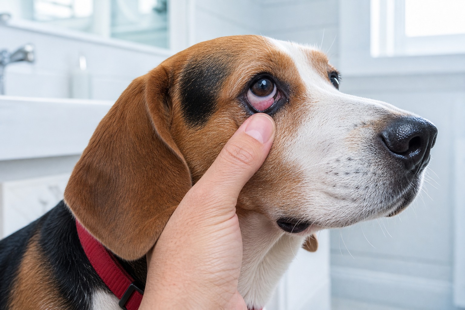
If you remember one thing about cherry eye, make it this: the gland stays. That single rule, insist on repositioning the gland rather than removing it, does more to protect your dog’s vision over its lifetime than anything else you will read here. It sounds like a small surgical detail. It is actually the difference between a dog whose eyes stay healthy and a dog on lifelong medication for chronic dry eye.
Everything else, how to spot cherry eye, why it happens, how fast to move, is in service of getting your dog to that one correct decision. Let’s start with recognizing it.
What cherry eye actually is
Here is something many people don’t realize: dogs have three eyelids, not two. The third eyelid rises from the inner corner of the eye and sweeps diagonally across to keep everything lubricated. Tucked into it is a tear gland that does a surprising amount of the eye’s work.
Cherry eye happens when that third eyelid gland slips out of its normal position and pops into view as a swollen red mass in the inner corner of the eye. The bulge can be constant, or it can come and go. It is not usually very painful, the signs are easy to spot, and treated quickly it typically leaves no lasting damage. Ignored, it can require surgery and lead to permanent problems like reduced tear production.
The honest part is that we still can’t always tell owners why that gland decided to slip. But what we can promise is that the sooner it’s back in place, and the more we protect the gland itself, the better that eye does for the rest of the dog’s life.
While any dog can develop cherry eye, it shows up most in certain breeds: American Cocker Spaniels, Basset Hounds, Beagles, Boston Terriers, English Bulldogs, Lhasa Apsos, Pekingese, Shih Tzus, and other flat-faced (brachycephalic) breeds. If you have one of these, it is worth knowing the signs in advance.
Signs to watch for
The signs tend to be visible and fairly distinct:
- A pink or red rounded bulge in the inner corner of the eye
- Irritation, including pawing or rubbing at the eye
- Trouble fully closing the eye
- Eye discharge
Move fast: what to do the day you see it
If you spot a red, swollen bulge in the inner corner of your dog’s eye, large or small, call your vet right away. Speed matters for a specific, evidence-based reason, not just general caution. The longer that gland sits out of place, the more the eye suffers downstream. Chronic prolapse can lead to conjunctivitis (pink eye) and ongoing discharge, and there is good evidence that correcting it early lowers a dog’s chance of developing low tear production later on.
That downstream risk is the whole reason to hurry. Reduced tear production, or dry eye, brings its own cascade: diminished vision, corneal ulcers that can become infected, and in the worst cases a ruptured eye. Dry eye usually responds to tear-stimulating therapy, but it often means lifelong daily medication. Catch cherry eye early and you sidestep most of that.
The one rule: reposition, never remove
Now the part that matters most. There are two philosophies a vet can bring to cherry eye, and only one of them is current.
The outdated approach is to simply cut the prolapsed gland out. It is fast, it makes the red bump disappear, and for decades it was common. It is also now discouraged by veterinary ophthalmologists, because that third eyelid gland produces roughly 30 to 50 percent of a dog’s tears. Remove it and you have traded a visible bump for a hidden, permanent deficit, dramatically raising the odds of lifelong dry eye. If a vet recommends removing the gland, that is your cue to ask about gland-sparing options or request a referral to an ophthalmologist.
The modern standard is to surgically reposition the gland back where it belongs and leave it intact. The most common gland-sparing method is the pocket technique (often called the Morgan pocket technique), where the surgeon tucks the gland into a small pocket of conjunctiva and closes it in. A related approach, tacking or tucking, anchors the gland back into position with a stitch. These procedures work well. Studies of the pocket technique report success rates in the mid-90s, commonly around 94 to 96 percent, with recurrence typically under 10 percent. If your dog’s cherry eye is intermittent, your vet may wait until it is in constant prolapse before operating, which is normal.
“Parents sometimes push for the quick removal because they just want the bump gone,” Dr. Park notes. “I understand the instinct, but I always slow that conversation down. You cannot give the gland back once it’s out. Repositioning takes a little more skill and a little more healing time, and it’s worth every bit of both.”
Treatment doesn’t always start with surgery, either. A topical anti-inflammatory is sometimes the first step, especially when local inflammation is feeding the prolapse. If that fails or the gland slips again, gland-sparing surgery is the next move.
Timing changes the outcome
One more evidence-based point on urgency. The risk of dry eye after surgery is highest in dogs whose gland was prolapsed for a long stretch before it was put back. In plain terms, the longer that red bump sits there untreated, the more it can cost your dog even after a successful repair. Spotting it early and acting quickly genuinely changes the result.
Can you prevent it?
Unfortunately, there are no known ways to prevent cherry eye. About 40 percent of affected dogs develop it in both eyes, usually before age two, so if one eye goes, stay alert for the other. The best thing you can do is stay attentive and bring any concern to your vet quickly. Caught early, cherry eye is at its most treatable, and the gland is at its easiest to save.
If your dog seems to have it, head to the vet promptly. Your regular vet may be able to diagnose and treat it on the spot. If not, ask for a referral to a veterinary ophthalmologist, exactly the specialist most likely to save the gland rather than remove it.
References
- Peña, María Teresa, and Marta Leiva. “Canine Conjunctivitis and Blepharitis.” Veterinary Clinics of North America: Small Animal Practice, vol. 38, no. 2, 2008, pp. 233–249.
- American College of Veterinary Ophthalmologists. “Cherry Eye: Should It Stay or Should It Go?” acvo.org.
- Cornell University College of Veterinary Medicine, Riney Canine Health Center. “Cherry Eye in Dogs.” vet.cornell.edu.
- Multiple clinical evaluations of the (modified) Morgan pocket technique for prolapse of the third eyelid gland in dogs, reporting success rates of roughly 94–96 percent with low recurrence.
Common questions
Should the gland be removed?
No. Removing the third eyelid gland is an outdated fix that raises your dog's lifelong risk of dry eye, because that gland produces roughly 30 to 50 percent of the eye's tears. The modern standard is to reposition and save the gland, not cut it out. If a vet recommends removal, ask about gland-sparing surgery or a referral.
Will cherry eye affect both of my dog's eyes?
Often, yes. About 40 percent of dogs who develop cherry eye in one eye go on to develop it in the other, usually before age two.
Can cherry eye be prevented?
There are no proven ways to prevent it. The best protection is catching it early and getting your dog to the vet while it is still easy to treat.
Not a substitute for your vet. Our guidance is educational. For urgent symptoms, contact a vet or emergency clinic right away. Editorial standards
Dr. Park is a board-certified emergency and critical care specialist. She reviews our health and safety content for clinical accuracy and contributes guides on recognising urgent conditions. Her focus is helping owners act quickly and correctly when it matters most.
Meet our experts ↗Keep reading
 Health & WellnessYour Dog Isn't Getting Lazy in Winter. Their Body Chemistry Shifted.Dr. Lena Park · 5 min
 Health & WellnessMud Won't Hurt Your Dog. What's Living In It Might.Dr. Priya Nair · 6 min
 Health & WellnessImodium for Dogs: The Risks, the Dosing, and Why Vets HesitateDr. Ravi Mehta · 6 min
 Health & WellnessThe Dog-Killing Liver Fluke Just Crossed Into California. The Map Is the Real StoryDr. Ravi Mehta · 5 min
 Health & WellnessThe Vet Specialty Nobody Mentions, Until Your Dog Won't Stop ScratchingDr. Amara Solis · 6 min
 Health & WellnessA Dog Who Keeps Shaking Their Head Is on a ClockDr. Priya Nair · 8 min