


Eye Gunk Is Normal. The Color and the Count Tell You When It Isn't.
Two questions sort almost every case of dog eye discharge: what color is it, and is it one eye or both? Here's the triage, the eight conditions behind it, and the red flags that mean go now.

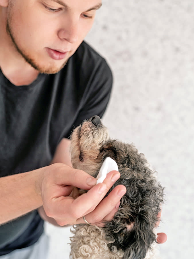
There’s a crust in the corner of your dog’s eye. There was one yesterday, too. You’ve been telling yourself it’s fine, and you’re probably right, because a certain amount of eye gunk is a completely normal byproduct of a working tear film.
But “probably right” is doing a lot of work there, and eyes are the one organ where waiting a week is a genuinely bad idea. A corneal ulcer can go from irritating to sight-threatening in days.
So skip the guesswork. There are two questions that sort almost every case, and you can answer both from across the room.
The two questions
Question 1: What color is it?
Tears exist to lubricate, protect, oxygenate, and feed the surface of the eye. When everything is working, the residue they leave is clear or slightly opaque, sometimes a grey-brown crust in the corner after sleep.
Color is a change in the content of the discharge, and it means something new is in there.
- Clear or opaque → normal tear residue, or simple irritation and excess tearing.
- Yellow or green → pus. That means infection is the leading concern, and it’s a same-day call.
- Thick, ropey, grey-green mucus → classically dry eye, where there isn’t enough watery tear to dilute the mucus component.
- Red or bloody → trauma, or a worsening problem inside the eye. Vet, now.
Question 2: Is it one eye or both?
This is the question almost nobody asks, and it’s arguably the more informative one.
Both eyes points toward something systemic or environmental: allergies, an infection moving through the body, dry eye, a viral respiratory illness, dusty air.
One eye points toward something local and mechanical: a foreign body, a scratch, an ulcer, a lid problem, an injury. And local mechanical problems are the ones with a clock on them.
A dog with sudden goop in one eye, squinting, is a same-day appointment. That combination is a corneal ulcer until proven otherwise, and untreated corneal ulcers can perforate.
Everything below fills in the detail behind those two questions.
The eight things behind excessive eye discharge
Conjunctivitis (“pink eye”)
Inflammation of the conjunctiva, the mucous membrane lining the inner eyelid and the whites of the eye. You’ll see redness, swelling, and discharge.
The important thing to understand is that conjunctivitis is a description, not a diagnosis. Something caused it: allergy, infection, trauma, or another eye disease underneath. Treatment depends on the cause, and typically involves reducing inflammation plus treating infection if it’s present. This is why “I have leftover eye drops from last time” is a bad plan; the wrong drops in the wrong eye can make things considerably worse.
Bacterial and viral infection
The big one here is canine infectious respiratory disease complex (CIRDC), commonly called kennel cough. It’s highly contagious, spread by respiratory droplets, direct contact, and contaminated objects like toys and bowls, and it’s usually caused by a combination of pathogens. The common culprits include:
- Canine parainfluenza virus
- Canine adenovirus type 2
- Canine influenza virus
- Canine respiratory coronavirus
- Canine herpesvirus-1
- Canine distemper virus
- Bordetella bronchiseptica
- Mycoplasma spp.
Dogs are often contagious for days before they show anything, which is how it moves so efficiently through boarding facilities and daycares. Symptoms include a dry, hacking cough, nasal discharge, eye discharge, sneezing, and lethargy. Most dogs recover with supportive care, but some progress to pneumonia, which brings fever and difficulty breathing and is an emergency.
If your dog has goopy eyes and a cough, you’re not looking at an eye problem. You’re looking at a respiratory problem that happens to include the eyes.
Dry eye (keratoconjunctivitis sicca, or KCS)
The classic cause of thick, gluey, unrelenting eye boogers.
Tears aren’t just water. When the lacrimal glands don’t produce enough of the watery component, the mucus component is left undiluted, and the result is thick, mucoid discharge, red and inflamed eyes, and, over time, a cornea that can scar and pigment and lose vision.
In dogs, KCS is most often immune-mediated: the immune system attacks the tear glands. That matters because it means it’s treatable, it’s usually lifelong, and it’s not going to resolve if you just keep wiping.
It’s also easy to test for. A Schirmer tear test takes about a minute in the exam room: a small paper strip is placed in the eye and the vet measures how far tears wick up it. If your dog has chronic gluey discharge and nobody has run one, ask.
Allergies
Dogs can react to proteins in food or to environmental triggers like dust, mold, and pollen. Itchy skin is the most common presentation, but allergic dogs also get chronic ear infections and red, itchy, watery eyes. Almost always both eyes, and almost always alongside other signs.
Foreign bodies
Anyone who has had a single eyelash in their eye understands the disproportionate misery involved. Dogs get hair, grass seeds, plant awns, and general debris in their eyes, and the response is a flood of tears plus frantic pawing.
Flushing with a dog-safe sterile eye wash is safer than letting your dog rub, and rubbing is what turns a foreign body into a corneal abrasion. If you can’t flush it out easily, stop and go in. Grass awns in particular can migrate and cause real damage.
Corneal abrasions
A scratch on the cornea, the clear window at the front of the eye. Caused by foreign material, a sharp object, a scuffle, or, very often, the dog’s own paw. Treatment is typically medicated drops plus a cone to stop further self-injury, and the cone is not optional.
Corneal ulcers
An abrasion that has become an open wound.
Ulcers come from dry eye, injury, infection, and other eye disease. They can be sterile or infected, shallow or deep, simple and quick to heal or indolent and slow. Deep ulcers that aren’t healing sometimes require surgery, and a perforated ulcer can cost the eye.
This is the reason “wait and see” is the wrong instinct with eyes. A dog squinting with discharge in one eye needs a fluorescein stain, which is a two-minute test that lights up an ulcer, and the sooner it’s found the smaller the problem is.
Anatomical abnormalities
Some dogs are simply built in a way that causes chronic irritation and tearing.
- Entropion: the eyelid rolls inward, so lashes and facial fur rub the cornea, causing inflammation and constant tearing. Usually the lower lid. Usually corrected surgically. Predisposed breeds include Labrador Retrievers, Chow Chows, Great Danes, English Bulldogs, and Bloodhounds.
- Ectropion: the eyelid rolls outward, exposing and drying the conjunctiva, which leads to inflammation, more tear production, and a higher infection risk. Commonly seen in Saint Bernards, Spaniels, Bloodhounds, Great Danes, and Newfoundlands.
- Lagophthalmos: the lids don’t shut all the way, so the cornea never gets fully covered, dries out, and takes damage it wouldn’t otherwise. Shallow eye sockets are the usual cause, which is why it’s common in brachycephalic (short-nosed) breeds like French Bulldogs, Boston Terriers, and Pugs.
If your dog is one of these breeds and has always had weepy eyes, the anatomy is a strong candidate, and it’s worth a conversation about whether it’s causing damage rather than just mess.
Owners apologize for bringing an eye in, and I always tell them the same thing: an eye is the one place where you’re allowed to be dramatic. If I look and it’s nothing, that took five minutes. If you wait and it was an ulcer, we’re talking about surgery.
Cleaning it, safely
Cleaning discharge away is legitimately useful. It’s more comfortable for your dog, and it lets topical medication actually reach the eye instead of getting absorbed by a crust. Follow your vet’s instructions if you’ve been given any.
How to do it:
- Be gentle. The first job is not to make anything worse.
- Use soft supplies. Cotton balls, cotton rounds, or a washcloth moistened with warm water. Sterile saline eye wash works too, but often isn’t necessary.
- Avoid anything abrasive. No paper towels, nothing stiff, nothing with a sharp corner. You can scratch a cornea with a paper towel.
- Wipe gently, away from the eye. If the crust is stubborn, hold a warm, moistened cotton ball against it as a compress for a minute to soften it, then wipe.
- Use a fresh piece of cotton for each eye. If one eye is infected, you don’t want to move it to the other.
Safe home care for the mild version — occasional crust, clear discharge, eyes look bright and white, no injury, dog is otherwise well:
- Clean gently as above.
- Do not let your dog rub or paw at the eye. If they’re doing it, put an e-collar on and call your vet.
- If something’s in the eye, rinse with a dog-safe saline eye wash. Never use Visine, contact lens solution, or human medicated drops.
- Keep watching. If “one small booger this morning” becomes “red, cloudy eye with discharge,” you’re done with home care.
Red flags: go now
Any change to the eye deserves at minimum a phone call. These deserve an immediate visit:
- Sudden blindness
- A change in eye color
- Bleeding in the eye
- A change in eye size
- A bulging eye
- Obvious eye pain (squinting, holding the eye shut, shying away from touch)
- A persistently abnormal pupil, unusually large or small
- Cloudiness on the surface of the eye
What newer research adds
The biggest practical change in recent years is in how dry eye gets treated, and it’s worth knowing before you’re in the exam room.
The mainstays are topical cyclosporine and tacrolimus, both calcineurin inhibitors that suppress the immune attack on the tear glands and let tear production recover. They are not lubricants; artificial tears help with comfort but do nothing about the underlying disease.
Between the two, current veterinary ophthalmology practice increasingly favors tacrolimus for more advanced cases, with reported success at increasing tear production in roughly 70 to 80 percent of dogs with KCS. Cyclosporine remains the standard first-line option, commercially available as a 0.2% ointment, with compounded solutions at higher concentrations; tacrolimus is typically compounded at 0.02% to 0.03%.
There is also active research into adjunct treatments, including injections of platelet-rich plasma into the tear glands, being explored as a cheaper option or an add-on to topical immunosuppressants. That’s still emerging, not standard care, but it’s a sign the field is moving.
The message for owners is simple: chronic gluey eyes are a diagnosable, treatable condition, not a grooming issue. Ask for a Schirmer tear test, and if the number is low, know that there are real drug options and that starting early protects the cornea.
Bottom line
Occasional clear crust is a normal side effect of tears doing their job.
Anything else, run the two questions. Yellow, green, red, or thick and ropey means something new is in the discharge. One eye plus squinting means something mechanical is wrong, and that’s the version with a deadline.
Wipe gently, never with a paper towel, never with human drops, and call your vet sooner than feels necessary. Eyes don’t reward patience.
FAQs
Which eye problems show up most often in dogs? Dry eye (KCS), eye infections, trauma, cataracts, glaucoma, uveitis, cherry eye, and anatomical problems with the eyelids. Any change to a dog’s eyes deserves prompt veterinary attention.
How do you treat a dog’s goopy eye? By finding out what’s causing it. Treatment ranges from a simple flush to a short course of drops to lifelong medication, and the wrong drops can make an ulcer significantly worse. Get it looked at rather than guessing.
What is canine distemper? A contagious infectious disease affecting the respiratory, gastrointestinal, and nervous systems. It causes GI upset, goopy eyes, and seizures. The prognosis is guarded to poor, which is why vaccination is strongly recommended.
Is green discharge from a dog’s eye serious? Yellow or green discharge means pus, which points to infection. Treat it as a same-day call to your vet.
Can I use human eye drops on my dog? No. Do not use Visine, contact lens solution, or human medicated drops. A dog-safe sterile saline eye wash is the only thing you should put in your dog’s eye without veterinary direction.
References
- “Canine Infectious Respiratory Disease Complex (Kennel Cough).” American Veterinary Medical Association. https://www.avma.org/resources-tools/pet-owners/petcare/canine-infectious-respiratory-disease-complex-kennel-cough
- “Disorders of the Cornea in Dogs.” MSD Veterinary Manual. https://www.msdvetmanual.com/dog-owners/eye-disorders-of-dogs/disorders-of-the-cornea-in-dogs
- Gelatt, Kirk N. “Disorders of the Eyelids in Dogs.” MSD Veterinary Manual. https://www.msdvetmanual.com/dog-owners/eye-disorders-of-dogs/disorders-of-the-eyelids-in-dogs
- Ofri, Ron. “Diagnosis and Treatment of Dry Eye in Dogs.” dvm360, vol. 54, 17 Mar. 2021. https://www.dvm360.com/view/diagnosis-and-treatment-of-dry-eye-in-dogs
- Ferrari, B., et al. “Comparison of Topical 0.03% Tacrolimus and Homologous Injectable Platelet-Rich Plasma in the Treatment of Keratoconjunctivitis Sicca in Dogs.” Frontiers in Veterinary Science, 2023. https://pmc.ncbi.nlm.nih.gov/articles/PMC9967714/
Not a substitute for your vet. Our guidance is educational. For urgent symptoms, contact a vet or emergency clinic right away. Editorial standards
Dr. Mehta is a board-certified veterinary nutritionist who evaluates pet food formulations, ingredient quality, and the science behind dietary trends. He writes and reviews all nutrition content at The Pet Times, including our food rankings and feeding guides.
Meet our experts ↗Keep reading
 Health & WellnessWhen an Old Dog's Bark Goes Hoarse, It's Rarely Just a Sore ThroatDr. Lena Park · 7 min
 Health & WellnessA Warm, Dry Nose Tells You Nothing. Here's How to Actually Know Your Dog Has a Fever.Dr. Priya Nair · 6 min
 Health & WellnessA Dog Who Won't Drink Water Is a Clock, Not a QuirkDr. Lena Park · 7 min
 Health & WellnessYour Dog's Poop Is a Daily Health Report. Here's How to Read ItDr. Lena Park · 8 min
 Health & WellnessMichigan's 'Mystery Dog Illness' Was Parvo All Along. The Test Was the Liar.Dr. Lena Park · 7 min
 Health & WellnessBloat in Dogs: The Deadly Emergency You Can Largely Prevent Before It StartsDr. Nina Kohl · 11 min