


You Found a Lump on Your Dog. Here's the One Thing You Can't Do About It.
You can't diagnose a lump by feeling it, and neither can your vet. Learn what cysts and tumors actually are, which changes should send you in fast, and why a five-minute needle test beats weeks of worrying.

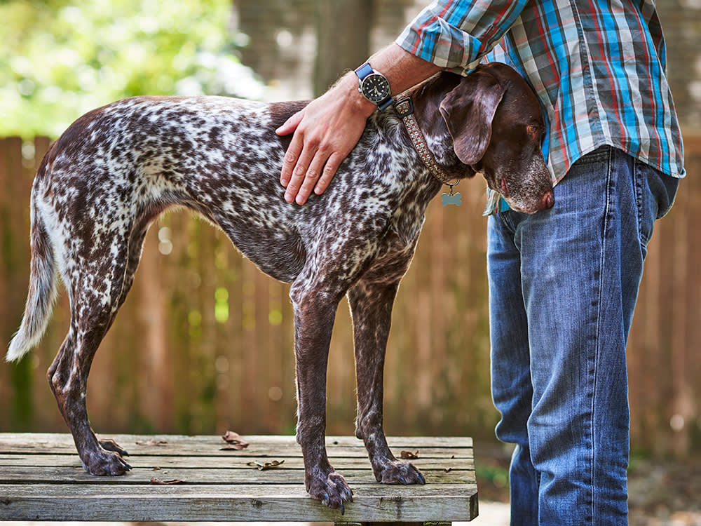
You are scratching your dog behind the ear, or running a hand down their side the way you have a thousand times, and your fingers stop on something that wasn’t there before. A bump. Your stomach drops. The word “cancer” arrives before any other thought does.
Here is the most useful thing to know in that moment, and it is going to feel unsatisfying: you cannot tell what the lump is by feeling it, and you are not supposed to be able to. Neither is your vet, not by touch alone. That sounds like bad news. It is actually the whole point, because it means the panic you feel and the danger the lump poses are two completely separate things, and the only tool that connects them is a quick test that most dogs barely notice.
Most new lumps are not cancer. Plenty are cysts, or benign growths that will sit there harmlessly for years. But a few are not, and the frustrating truth is that the harmless ones and the serious ones can feel identical under your hand. So the goal of this piece is not to help you diagnose the bump yourself. It is the opposite: to talk you out of trying, and to show you exactly which changes should move you from “keep an eye on it” to “call today.”
What a cyst actually is
A cyst is a closed sac, sitting in or just under the skin, that has filled up with something it shouldn’t be holding. Press on one and it can feel like a tiny water balloon. What’s trapped inside is usually watery fluid, a semi-solid paste, or keratin, the same tough protein your dog’s skin and nails are built from, here packed to a consistency somewhere between putty and toothpaste. The sac stays sealed unless it ruptures, and plenty of cysts are harmless, painless, and can be left alone indefinitely.
They form because something got blocked or trapped. A few common types tell the story:
Follicular (or epidermoid) cysts build up around a hair follicle and also fill with keratin; the usual spots to feel one are along the back, the sides, and the legs. Apocrine sweat gland cysts are rarer, tend to be softer and more fluid-filled, and turn up more in certain breeds. Sebaceous cysts, probably the type you’ll meet most, start when an oil gland plugs up; they’re typically round, slow to change, and can weep a thick pale material if they burst. And a false cyst isn’t really a cyst at all: it’s usually a seroma, a pocket of fluid that pools beneath the skin after an injury or surgery, with no true sac lining it.
None of that has much to do with cancer. The usual culprits behind a cyst are mundane: a plugged follicle, an oil gland working overtime, a small skin injury, an inherited tendency in some breeds, inflammation driven by allergies, or a lingering skin infection. Dogs with oily coats, repeat skin infections, or allergies tend to develop them more.
A cyst typically comes on slowly, feels smooth and rounded, and shifts a little under the skin when you nudge it with a fingertip. It won’t hurt unless it becomes infected, it may puff up and settle down over weeks, and a ruptured one drains thick fluid that’s white or yellowish. One thing worth flagging: if a cyst your vet already identified suddenly turns red, painful, swollen, or starts draining, that’s a sign it may be infected and deserves a recheck.
What a tumor actually is
A tumor is a mass of abnormal cells caused by cell division that got stuck in the “on” position. And here is the word people trip over: tumor does not mean cancer. Most tumors are benign, meaning they grow slowly and stay put. Malignant tumors, the ones we actually mean by cancer, can invade nearby tissue and spread elsewhere in the body. You cannot tell which is which by looking or feeling, full stop.
The common ones vary wildly, which is exactly why touch fails you. Take the two that matter most for this article, because they are the pair most often confused. A mast cell tumor is malignant, and it ranks among the skin cancers vets see most in dogs. It is also a shape-shifter: it can be tiny or bulky, hard or yielding, raw-looking or covered in skin that appears perfectly ordinary. A lipoma, by contrast, is a benign lump of fat, the kind that gives under your fingers and slides around, and it turns up constantly in dogs past middle age. Here is the problem: a mast cell tumor can pass for a lipoma under your hand. That single fact is the reason this article exists.
The rest of the roster is just as unhelpful to a fingertip. A histiocytoma is benign, a young dog’s lump, red and raised and quick to appear, and it commonly disappears without treatment. A melanoma may be benign or malignant, and it favors the skin, the nail beds, and the mouth. A soft tissue sarcoma sits at the other extreme: cancerous, capable of pushing into nearby tissue, and yet often so slow-growing that it lulls you.
The root cause is almost always a genetic mutation, and the things that raise the odds are largely out of your hands: getting older, inflammation that never resolves, the breed your dog happens to be, hormones, exposures in the environment, and a shifting immune system. A specific cause is rarely identifiable, and it is hardly ever something a pet parent did.
A tumor that’s causing problems tends to announce it. It might hurt, or throw off your dog’s gait if it sits somewhere awkward. It might break open at the surface or bleed. It might shift in size, shape, or shade, put on volume fast, or sit hard and anchored rather than sliding under your fingers. But — and this is the whole problem — plenty of cancerous lumps feel perfectly innocuous, and plenty of harmless ones feel alarming.
The comparison that matters, and its asterisk
Line the two up and a pattern does emerge. Cysts lean slow, soft, sac-like, and content to stay put; they slide, they’re usually harmless, and they can burst and weep. Tumors lean solid, and they’re the less predictable of the pair: harmless or dangerous, quick or creeping, sometimes anchored or lumpy to the touch, and not given to draining.
That table is real, and it is also a trap if you lean on it too hard. The pattern describes what’s typical, not what’s certain. The single most important sentence in this entire article is the one that pattern can’t override: fingertips cannot tell you what a lump is. Not yours, and not those of a vet who has felt ten thousand of them. Answers come from tests.
The test you’re afraid of is the easy part
This is where the fear usually lives, and it shouldn’t. Finding out what a lump is rarely means surgery. It usually starts with a fine needle aspirate, or FNA: a quick needle stick that pulls a few cells to look at under a microscope. Most dogs tolerate it about as well as a vaccine, it often happens in the same visit, and it’s frequently all it takes to say “this is fat, stop worrying” or “this needs a closer look.”
When the needle isn’t enough, a biopsy removes a small piece of tissue for the lab and gives a definitive answer. Imaging like X-rays or ultrasound comes in when your vet needs to check for internal masses or see whether something has spread.
From there, treatment follows the diagnosis, not the other way around. A benign cyst or tumor may simply be watched. An infected cyst usually needs antibiotics or anti-inflammatories. Masses that are painful, infected, growing fast, or malignant may need surgical removal. And a malignant tumor may call for oncology care such as chemotherapy, radiation, or targeted therapy. The through-line across all of it: the earlier you catch a lump and get it checked, the smaller and simpler the fix, and the better the odds.
The mistake I see isn’t people overreacting to lumps, it’s people negotiating with them. They decide it’s probably fat, they wait to see if it grows, and they hand a fast-moving tumor a head start it didn’t need. A needle sample takes five minutes and ends the guessing. I would always rather tell you it’s nothing than meet a lump six months too late.
What’s new since this was written
There’s a genuinely encouraging development for one of the scarier lumps on this list. For dogs with certain non-metastatic mast cell tumors, the FDA has approved an injectable drug called Stelfonta (tigilanol tiglate) that is given directly into the tumor rather than cut out. In the company’s clinical data, it cleared about 75 percent of tumors after a single treatment and roughly 87 percent after one or two, which gives some dogs a non-surgical option that didn’t exist a few years ago.
It isn’t right for every tumor or every location, and it doesn’t change the first move at all: a mast cell tumor still has to be identified by a needle sample before anyone can treat it. But it’s a real example of why catching a lump early matters more than ever. The more options that exist for a small, contained tumor, the more you gain by not letting it grow. Interestingly, the same drug was granted orphan-drug status for a type of human cancer in early 2024 and is now working through human trials, so this is an area moving quickly.
The one habit worth building
You can’t prevent every lump, but you can change how early you find them. Once a month, run both hands slowly over your entire dog, not to diagnose anything but simply to learn what “normal” feels like for their body, so a new bump stands out the next time. Keep them at a healthy weight, manage allergies, get skin infections looked at rather than waiting them out, and keep up with regular vet exams. And don’t dismiss a lump just because it’s small or hasn’t changed. Small is the best time to check, not a reason to skip it.
Finding a lump is genuinely frightening, and the fear is real even when the lump isn’t dangerous. But the fear and the danger are not the same thing, and one quick test is what tells them apart. Guessing keeps you stuck in the scary part. Testing gets you out of it, usually with good news, and occasionally with a head start that matters.
This is a topic where the details are individual to your dog, so treat this as background rather than a substitute for a hands-on exam. If you’ve found something new, growing, or changing, your vet is the fastest way from worry to an answer.
References
- Adedeji, Adeyemi O., et al. “Cytologic Features of Cutaneous Follicular Tumors and Cysts in Dogs.” Veterinary Clinical Pathology, vol. 46, no. 1, Mar. 2017, pp. 143–150.
- Dobson, Jane M. “Breed-Predispositions to Cancer in Pedigree Dogs.” ISRN Veterinary Science, vol. 2013, 2013, pp. 1–23.
- Garrett, Laura. “Canine Mast Cell Tumors: Diagnosis, Treatment, and Prognosis.” Veterinary Medicine: Research and Reports, vol. 5, Aug. 2014, p. 49.
- U.S. Food and Drug Administration. “FDA Approves First Intratumoral Injection to Treat Non-Metastatic Mast Cell Tumors in Dogs.” FDA CVM Updates.
- Musser, M., et al. “Response to Tigilanol Tiglate in Dogs with Mast Cell Tumors.” Journal of Veterinary Internal Medicine, 2024.
Common questions
Do all lumps on dogs need to be removed?
No. Many cysts and benign tumors like lipomas can simply be monitored once they've been identified. Removal is reserved for lumps that are painful, infected, growing quickly, interfering with movement, or confirmed to be malignant.
How fast should I get a new lump checked?
Sooner is always better. A lump that is new, growing, changing shape or color, ulcerated, bleeding, or bothering your dog should be seen promptly. Early, small masses are almost always easier and cheaper to treat than ones left to grow.
Not a substitute for your vet. Our guidance is educational. For urgent symptoms, contact a vet or emergency clinic right away. Editorial standards
Dr. Kohl is a board-certified veterinary dentist with a particular interest in preventive oral care. She reviews all dental health content at The Pet Times and contributes practical guides on home dental routines, professional cleaning, and oral disease prevention.
Meet our experts ↗Keep reading
 Health & WellnessBone Cancer in Big Dogs: Don't Wait Out the LimpDr. Mara Chen · 7 min
 Health & WellnessMichigan's 'Mystery Dog Illness' Was Parvo All Along. The Test Was the Liar.Dr. Lena Park · 7 min
 Health & WellnessYour Dog Eating Dirt Is Usually Boredom. Sometimes It's Their Blood.Dr. Mara Chen · 7 min
 Health & WellnessYour Dog's Resting Breathing Rate Is an Early-Warning System. Here's How to Read It.Dr. Lena Park · 6 min
 Health & WellnessOne Mosquito Can Put a Worm in Your Dog's Heart. Here's How to Stop It.Dr. Ravi Mehta · 8 min
 Health & WellnessYour Dog's Nose Predicts Their Lifespan. Nobody Wants to Hear It.Dr. Mara Chen · 7 min